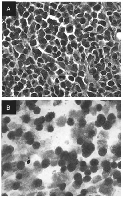
FIGURE 2.

(A) Histopathology of skin biopsy showing anaplastic lymphoid cells with pleomorphic nuclei in mitosis (hematoxylin & eosin stain, 200×). (B) Cytology of vitreous biopsy showing atypical lymphoid cells containing large irregular nuclei, few reactive lymphocytes, and macrophages (Giemsa stain, 400×).
