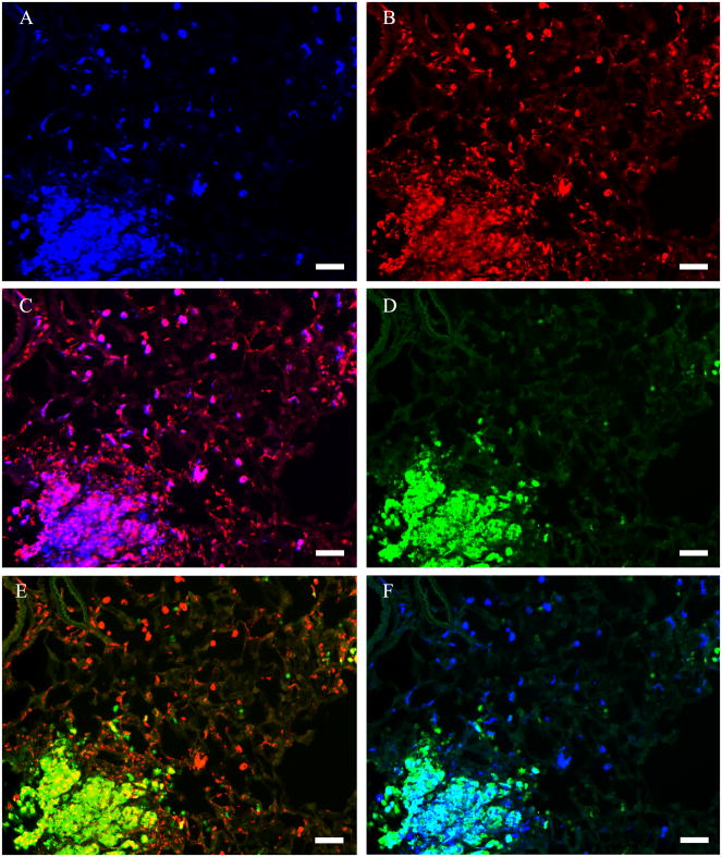
Figure 6. Identification of cells expressing arginase I and iNOS in silica-treated rat lung.
Results are shown for rats treated with 5 mg silica/100 g BW. The tissue section was stained with anti-arginase I (blue), anti-iNOS (green) and the macrophage marker ED-1 (red). (A) Identification of arginase I-positive cells. (B) Identification of macrophages with the macrophage marker ED-1. (C) Identification of arginase I-positive macrophages by colocalization of immunoreactivity for arginase I and ED-1 (pink). (D) Identification of iNOS-positive cells. (E) Identification of iNOS-positive macrophages by colocalization of immunoreactivity for iNOS and ED-1 (yellow). (F) Identification of cells expressing both arginase I and iNOS by colocalization of immunoreactivity for arginase I and iNOS (turquoise). Photomicrographs are representative of multiple sections examined for each of 8 silica-treated rats. Scale bar = 50 μm.

