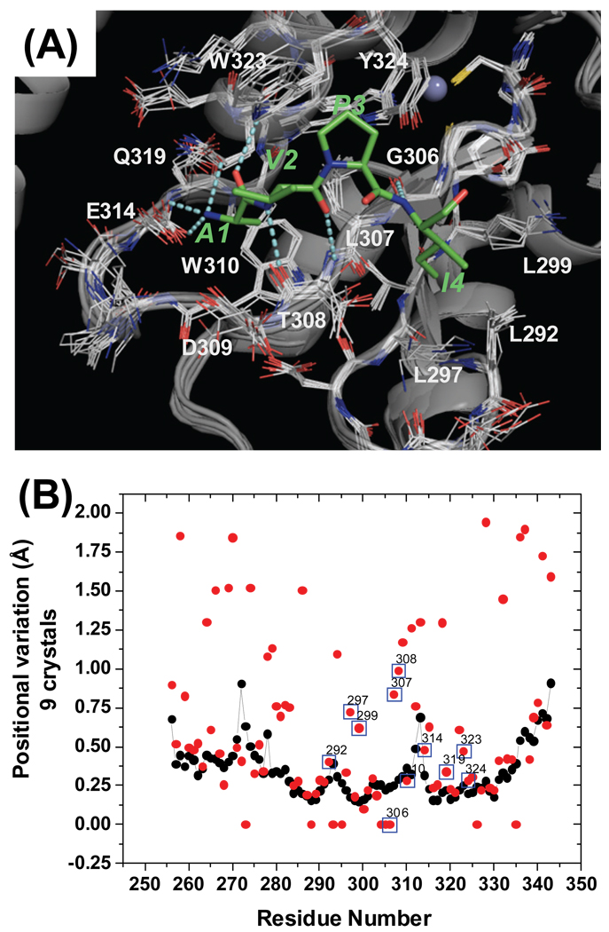
Figure 2.
(A) Structure of the XIAP BIR3 - AVPI complex and alignment of the nine ligand bound XIAP BIR3 crystal structures. Residues around the binding site (L292-Y324) are shown as line structures. The AVPI peptide is colored in green. Residues of XIAP BIR3 which directly interact with AVPI are labeled. (B) Per residue positional variation of XIAP BIR3 calculated from the alignment of nine structures. Black points correspond to backbone atoms, red points to side chain atoms.

