
Figure 1.
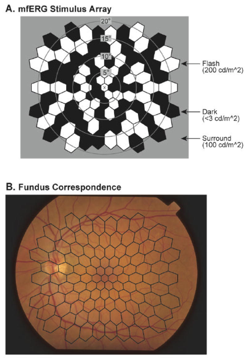
The multifocal stimulus. A: The stimulus array is comprised of 103 hexagonal elements that are scaled with retinal eccentricity. The X in the center is the fixation target. The stimulus elements are modulated pseudorandomly between black (< 3 cd/m2) and white (200 cd/m2) according to an m-sequence. B: Spatial correspondence between the stimulus array and the retina is shown. The fundus photograph is of the left eye of a diabetic patient with no retinopathy.