Fig. 3.
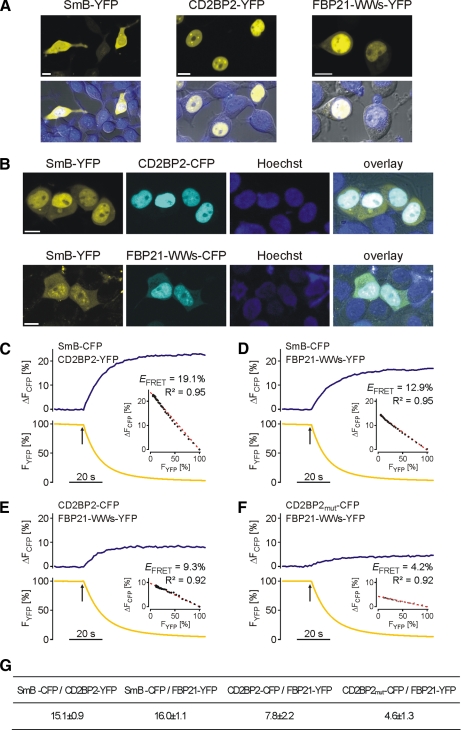
Localization and FRET analysis of SmB-associated proteins. A, confocal laser scanning images of YFP fusion proteins of CD2BP2/52K, SmB, and FBP21. The respective proteins were transiently expressed in HEK293T cells and imaged with a confocal laser scanning microscope (LSM510-META) equipped with an α-Plan Fluar 100/1.45 objective microscope. Bar, 10 μm. B, co-expression of YFP-fused SmB with CD2BP2/52K-CFP or CFP-tagged FBP21 tandem WW domain construct. Images were recorded as described in A. The SmB-YFP was tracked to the nucleus by CD2BP2/52K-CFP or CFP-tagged FBP21-WWs. C–F, analysis of fluorescence energy transfer of a single experiment between SmB-CFP and CD2BP2-YFP (C), SmB-CFP and FBP21-YFP (D), CD2BP2-CFP and FBP21-YFP (E), and CD2BP2-CFPmut and FBP21-YFP (F). During acceptor photobleaching the donor (CFP) fluorescence signal increases whereas the acceptor (YFP) fluorescence decreases. FRET efficiency was calculated by regression analysis of the various intensities obtained during photobleaching of YFP. CD2BP2-CFPmut denotes a GYF mutant (W8R/Y33A) of CD2BP2 that is devoid of PRS binding. G, FRET efficiencies of energy transfer. Data represent mean ± S.E. of a minimum of four independent measurements.

