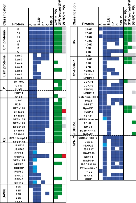
Table I. Proteins found in the pulldown experiments compared with known spliceosomal complexes.
The presence of proteins in the spliceosomal complexes A (33), B (32), BΔU1, B*, C and 35 S U5 (47) is indicated by blue squares. Light blue squares represent proteins only found once in four preparations of the spliceosomal B complex (31). Proteins identified by our pulldown experiments using GST-GYF in the presence or absence of PD1 peptide (GYF +/− PD1) using a combination of GST-tagged WT and mutant GYF domain (GYF/mutant-GYF) or using GST-tagged U5-15K in the presence or absence of PD1 peptide (U5-15K +/− PD1) are indicated in the last three columns. Green squares represent labeled proteins, red squares represent unlabeled proteins, and gray squares represent a mixed state. SmB could not be identified in pulldowns with GST-GYF according to the selection criteria. Additional evidence for selective binding from other experiments (Fig. 4C) led to the classification of SmB protein as labeled, and it is represented by green, hatched squares. Proteins are grouped according to subcomplexes. Hashed lines separate true components (upper part) from complex-related proteins (lower part).

