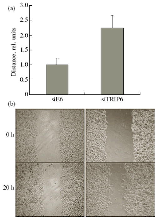
Figure 2.
Migration of A549 cells in the wound-healing test. A linear wound in a cell monolayer was inflicted with a plastic pipette tip; the cells were allowed to migrate for 20 h and were photographed. (a) Cell migration rate was normalized to the control (siE6) (M ± SD). The results of three independent experiments performed in triplicate are shown. (b) A typical “wound” at the beginning of experiment (upper row) and 20 h later (lower row), phase contrast microscopy.

