
Figure 7. Mammary Epithelia Are in a Multilayered State during Morphogenesis.
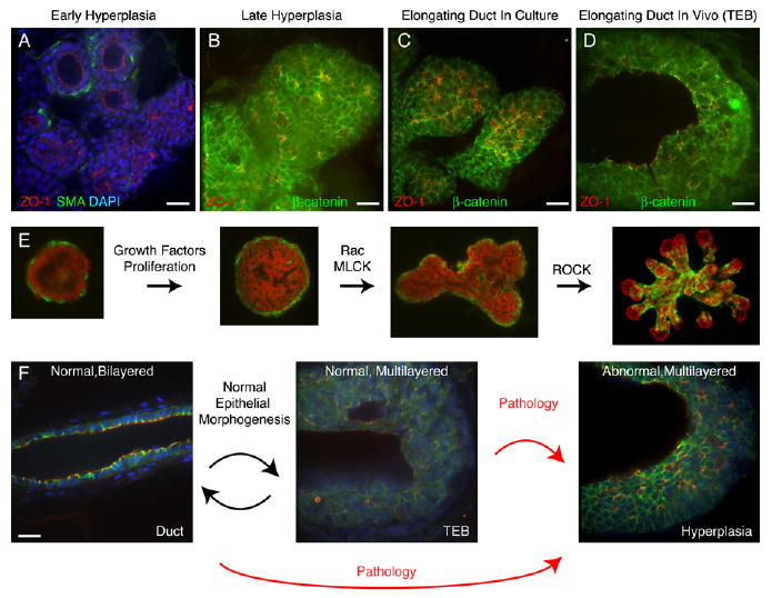
(A) Early hyperplasias from the MMTV-PymT mouse model lose myoepithelial (ME) coverage and transition into a multilayered epithelial organization.
(B) Late hyperplasias from the MMTV-PymT mouse model have a multilayered epithelial organization and consist of incompletely polarized cells, with β-catenin on all lateral surfaces.
(C and D) Normal (C) FGF2-treated ducts in culture and (D) terminal end buds in vivo are multilayered and consist of incompletely polarized cells, with β-catenin on all lateral surfaces.
(E) Schematic representation of epithelial morphogenesis in culture.
(F) During normal morphogenesis, mammary epithelium transiently reorganizes into a highly proliferative, low-polarity, multilayered state and then returns to a quiescent, simple epithelial state. Aberrant entry into, or failure to exit from, this morphogenetically active state may facilitate tumor initiation or progression.
Scale bars are 20 μm.