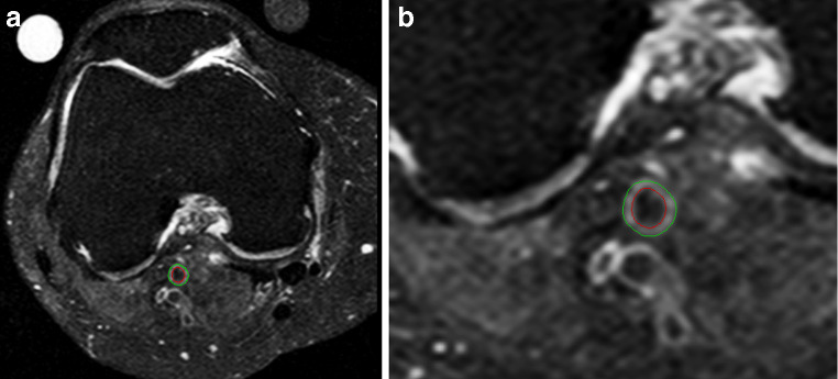
Fig. 1.
a Axial fat-suppressed spin echo image of the knee after semi-automatic vessel wall thickness quantification in a patient with generalized OA. Note the osteophytes at the femoral condyle (arrow). b Detail of the semi-automatic vessel wall thickness quantification of the luminal and outer wall boundaries of the vessel wall

