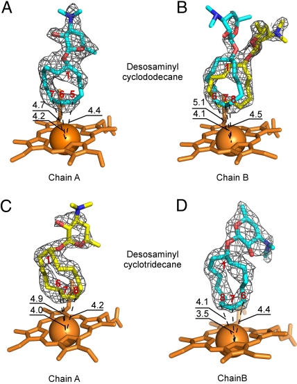
Fig. 4.
Multiple binding modes of desosaminyl cycloalkanes. Orientations of structure 6 in the active site of PikCD50N (A) in chain A and (B) in chain B, and orientations of structure 8 in the active site of PikCD50N (C) in chain A and (D) in chain B, as defined by the fragments of the electron density map (gray mesh) contoured at 0.8 σ are shown. In (B), structure 6 is docked in the flipped-over orientations, allowing hydroxylation on the both sides of the ring. In (C) and (D), structure 8 is in flipped-over orientations. Heme is shown in orange. Oxygen atoms are in red, nitrogen in blue, iron in orange (shown as a Van der Waals sphere). Atoms of the cycloalkane ring are labeled in red. Distances are in Angstroms. Images are generated using PYMOL.

