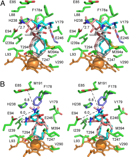
Fig. 5.
Desosaminyl cycloalkane binding sites. (A) Stereoview of the PikCD50N binding site with the three superimposed structure 6 conformers highlighted (gray) in chain A and (pink and cyan) chain B, surrounded by the chain-A amino acid side chains within 5 Å plus E85 (green). E246 of chain B is highlighted in ice blue. For clarity, V242 was omitted from the drawing. (B) Stereoview of the PikCD50N binding site with two superimposed structure 8 conformers highlighted (pink) in chain A and (cyan) chain B, surrounded by the chain A amino acid side chains within 5 Å plus E85. F178 of chain B is highlighted in ice blue. Heme is shown in orange. Oxygen atoms are in red, nitrogen in blue, iron in orange (shown as a Van der Waals sphere). Distances between tertiary amine and carboxylic groups are in Angstroms. Lowercase “a” in the residue label indicates that alternative conformations are shown.

