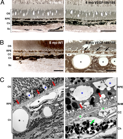
Fig. 1.
Abnormal choroidal vasculature and RPE defects in aged VEGF188/188 mice. (A and B) One-micrometer-thin epon sections of 4- and 8-month-old wt (right panel) and VEGF188/188 (left panel) mice were stained with para-phenylenediamine for light microscopy (A and B) or were visualized by TEM (C). (A) No changes in the organization of the retina layers were observed in 4-month-old VEGF188/188 mice. (B) An 8-month-old, VEGF188/188 mouse eye displays major abnormalities in the RPE and choroidal vessels. Larges vacuoles were observed within the RPE (arrows). The choroidal vessels appear enlarged, and the CC is replaced by discontinuous vessels wrapped by pericytes (asterisk). (C) Electron micrograph of the mid-periphery of an 8-month-old VEGF188/188 mouse retina. Note the significant enlargement of the choroidal vessels (black asterisk). RPE cells displayed large vacuoles containing membranous debris (blue asterisk). Regression of the RPE basal infoldings is associated with accumulation of materials (red arrow). Higher magnification (right) showing discontinuous collagen and elastin layers in the BrM (green asterisk) and a regressing CC vessel with an entrapped erythrocyte (green arrow). [Scale bar, 50 μm (A); scale bar, 20 μm (B); scale bar, 2 μm (C).] OS, outer segment; BrM, Bruch's membrane; CC, choriocapillaris; Ch, choroid; Sc, sclera.

