
Figure 1.
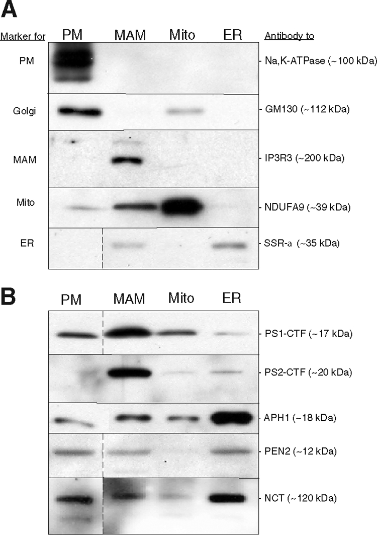
Western blot analysis of subcellular fractions of mouse brain. Thirty μg of total protein were loaded in each lane. A: Localization and predicted molecular masses of the indicated polypeptides were determined using the antibodies listed at right (see text). PM, plasma membrane. B: Fractions were probed using the indicated antibodies against PS1 (Calbiochem PC267) and PS2 (Cell Signaling 2192) and to other components of the γ-secretase complex. In the blots shown here, the intensity of both the PS1 and the PS2 signals in MAM was enriched ∼eightfold over that in the ER. In some blots, data represent nonadjacent lanes taken from a single blot; dividing lines indicate where lanes were pasted together.