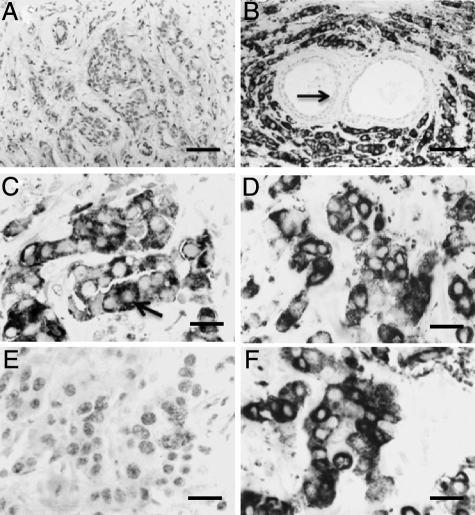
Figure 1.
Immunocytochemical staining of primary carcinomas by AGR2 mAb. A: Section of an invasive carcinoma showing no immunocytochemical staining for AGR2. B: Section of an invasive carcinoma showing strong positive staining for AGR2 (+++). The normal glandular tissue is unstained (arrow). C: Section of an invasive carcinoma at higher magnification showing positive staining for AGR2 (+++) in secretory granules (arrow). D–F: Serial sections of the same carcinoma incubated with D, mAb to AGR2 showing positive cytoplasmic staining (+++); E, mAb to AGR2 plus 700 μg/ml recombinant AGR2 showing no staining; or F, mAb to AGR2 plus 700 μg/ml recombinant AGR3 showing no diminution of positive staining (+++) over that in D. Magnification, A and B, ×220; C, ×685; D–F, ×545. Bars, A and B, 50 μm; C, 15 μm; D–F, 20 μm.

