Figure 2.
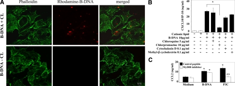
Cationic lipid enhances the uptake of B-DNA in GEnC. A: GENCs were exposed to 5 μg rhodamine-labeled B-DNA in the presence or absence of cationic lipid (CL) for 2 hours. Intracellular uptake was detected by confocal microscopy and appears as red staining inside GEnCs. FITC-labeled phalloidin was used to mark the cytoskeleton of GEnCs and appears as green staining. Note that the intracellular uptake of B-DNA depended on the presence of CL. Images are representative for three independent experiments. Original magnification ×400. B: GEnCs were stimulated with either CL alone or B-DNA/CL in the presence or absence of chlorpromazine, methyl-β cyclodextrin, cytochalasin D, or chloroquine as indicated. CXCL10/IP-10 was determined in supernatants after 24 hours by ELISA. Data are means ± SEM from three experiments each analyzed in duplicates. *P < 0.05. C: GEnC were preincubated with 100 μmol/L MyD88 homodimerization inhibitory peptide or control peptide for 24 hours and then stimulated with either CL alone or B-DNA/CL. CCL2/MCP-1 were determined in supernatants after 24 hours by ELISA. Data are means ± SEM from three experiments analyzed in duplicate. *P < 0.05 vs. medium, **P < 0.05 vs. control peptide.

