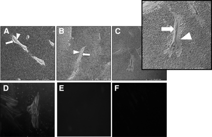
Figure 8.
Effect of HAS2 overexpression on pericellular HA coat assembly and phenotype after TGF-β1 in aged dermal fibroblasts. Patient-matched young (A and D) and aged (B, C, E, and F) dermal fibroblasts were transfected either with pCR3.1 alone (A, B, D, and E) or HAS2-pCR3.1 (C and F). Forty-eight hours after transfection, cells were incubated in serum-free medium containing 10 ng/ml TGF-β1 for 72 hours. The pericellular HA coat (A–C) was visualized by addition of formalized horse erythrocytes. Arrows indicate the cell body; arrowheads show the extent of the pericellular matrix. Images are representative of dermal fibroblasts from three patient donors. Original magnification, ×200. After stimulation the cells were fixed, and α-SMA was visualized by immunohistochemistry (D–F). Slides were mounted in Vectashield fluorescent mountant and viewed under UV light. Original magnification, ×100.

