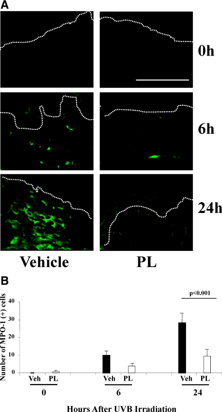
Figure 3.
A: Representative images of immunostained (green fluorescence) MPO-1 positive (+) cells (neutrophils) in mice treated with PL or vehicle at 0, 6, and 24 hours after UV radiation. Please note that by 48 and 72 hours after UV irradiation the number of MPO-1+ cells were negligible in PL-fed and vehicle-fed mice (data not shown), which did not permit quantification of MPO-1+ cells to allow comparison of the effects. B: Graphic representation of the number of MPO-1+ cells per 200 linear μm of epidermal length showed threefold significant decrease (N = 5 per treatment group, of six to eight visual fields/mouse) of MPO-1+ cells in PL-fed mice 24 hours after UV irradiation. Dermo-epidermal junctions (D-E) were identified by PI nuclear staining (see Supplemental Figure S3 at http://ajp.amjpathol.org) and are outlined by white dotted lines. Scale bar = 200 μm.

