Figure 5.
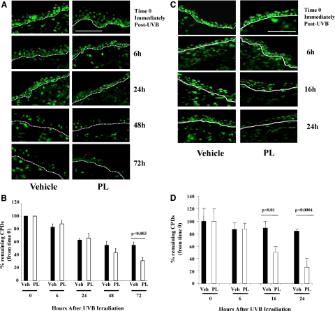
A: Representative images of immunostained (green fluorescence) CPDs+ cells in Xpc+/− mice treated with PL or vehicle at 0, 6, 24, 48, and 72 hours after UV radiation. In vehicle-fed Xpc+/− mice, CPD positivity persisted up to 72 hours post-UVB, whereas by 72 hours there was noticeably less detectable CPDs remained in PL-fed Xpc+/− mouse skin (in epidermis as well as dermis). Percentage of remaining detectable CPDs were determined as the ratio of the CPD+ nuclei at that time compared with that immediately after irradiation (time 0). D-E junctions are outlined by dotted lines. Scale bar = 200 μm. B: Graphic representation of CPD+ nuclei per 200 linear μm of epidermal length showed a statistically significant decrease of percent remaining CPDs in PL-fed Xpc+/− mice at 72 hours. C: Representative images of immunostained (green fluorescence) CPDs+ cells in wild-type mice treated with PL or vehicle at 0, 6, 16, and 24 hours after UV irradiation. In vehicle-fed wild-type mice more than 80% CPD positivity persisted up to 24 hours post-UVB, whereas by 16 and 24 hours there was noticeably less detectable CPDs remained in PL-fed wild-type mouse skin (in epidermis as well as dermis). Percentage of remaining detectable CPDs were determined as the ratio of the CPD+ nuclei at that time compared with that immediately after irradiation (time 0). D-E junctions are outlined by dotted lines. Scale bar = 200 μm. D: Graphic representation of CPD+ nuclei per 200 linear μm of epidermal length showed statistically significant decrease of percent remaining CPDs in PL-fed wild-type mice (clear bars) at 16 and 24 hours.

