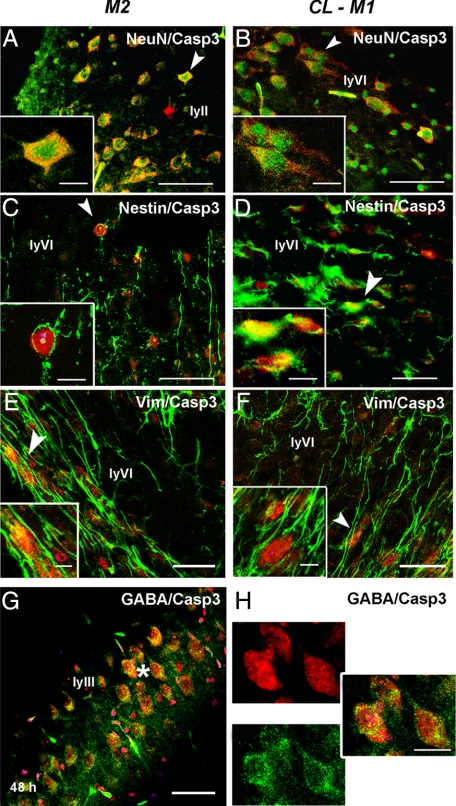
Figure 3.
Unilateral transient CCA occlusion mainly triggers casp3 cleavage in neurons and undifferentiated cells in the P7 rat brain. Contralateral cortex from uni-CCAo (model M2) and MCAo + tCCAo (model M1) animals contain most cleaved casp3 colocalized with neurons (NeuN) (A, B), nestin- (C, D), and vimentin (E, F) positive cells Arrows indicate location magnified in insets. Co-localization of cleaved casp3 was observed in GABA-immunostained neurons in cortical layer III (ly III in G, and enlarged panels in H, indicated by the asterisk in G). Cleaved casp3 labeling is shown in red, whereas NeuN, nestin, vimentin and GABA markers are shown in green. Scale bar represents 50 and 20 μm (enlarged panels).

