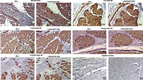
Figure 10.
Cripto-1 cardiac expression in an animal model of MI. Immunohistochemistry for CR-1 and α-sarcomeric actin in serial heart tissue sections of a porcine model of myocardial ischemia-reperfusion. Heart tissue samples were collected after 2 hours, 3 days, 10 days, and 2 months of induced MI. A normal heart sample was harvested from a healthy pig. Red arrows in the normal heart are pointing to some of the cells that show positive staining for CR-1. Negative controls were obtained by omitting the primary antibodies only. Scale bar = 50 μm. Original magnifications, ×20.

