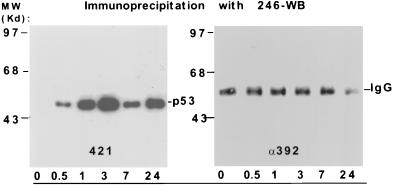
Figure 3.
Western blot analyses of p53 levels and phosphorylation patterns after γ irradiation. F9 cells were exposed to 7 Gy of γ radiation and harvested at 0, 0.5, 1, 3, 7, and 24 hr after exposure. After immunoprecipitation with Pab246, the transferred gels were probed with PAb421 (421) or α-392 antibodies. Molecular weight markers are indicated at left. The p53 bands in the 421 blot were detected with a 15-sec exposure. The IgG bands (at a higher molecule weight than p53) are present in the α-392 blot and were detected with a 15-min exposure (no bands were observed with a 15-sec exposure) used to increase the sensitivity of p53 detection. No phosphorylated form of p53 serine-389 was observed.

