
Figure 3. IL-6 protects MSCs from serum starvation-induced apoptosis.
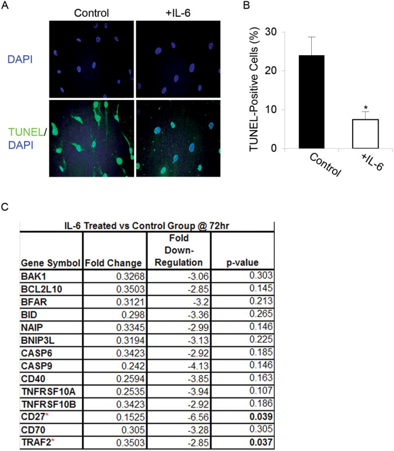
(A) TUNEL: Apoptosis was evaluated using TUNEL staining in undifferentiated MSCs grown in serum-free media for 72 h with or without IL-6. IL-6 treated populations exhibited fewer apoptotic cells compared to untreated controls. DAPI staining revealed IL-6 treated cells had smaller, more pyknotic nuclei and TUNEL staining was more intense in MSCs serum-starved and treated with IL-6. Magnification: 20X. (B) Cell counts: For each condition, total numbers of TUNEL-positive cells were counted and a ratio of the means was calculated. The mean ratio of apoptotic cells in serum-free conditions to apoptotic cells in IL-6 supplemented conditions was 3.5: 1 (t-test: * p < 0.05). (C) Microarray gene expression analysis: Expression levels of pro-apoptotic genes decreased upon treatment with IL-6 for 72 h, only significantly for CD27 and TRAF2.