
Figure 1.
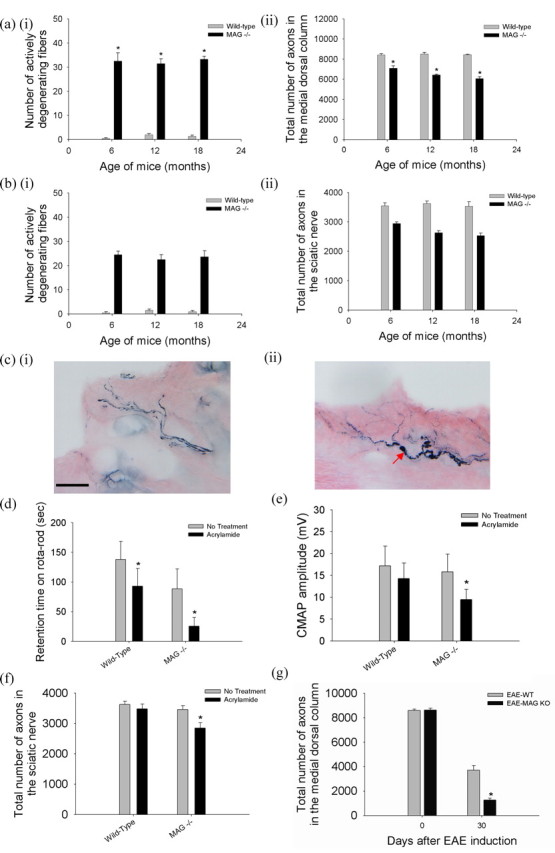
MAG promotes axonal stability in vivo. C5 spinal cords, sciatic nerves proximal to the bifurcation, and distal tibial nerves at the ankles were harvested from groups of five mice at 6, 12, and 15 months of age. Every myelinated fiber was counted in easily defined regions of the CNS and PNS as shown in supplemental Figure S1 (available at www.jneurosci.org as supplemental material). a, In the medial dorsal columns at C5 in wild-type mice, there were few degenerating fibers (ai), and axonal numbers remained constant between 6 and 15 months of age (aii). In contrast, MAG−/− mice had a high number of active Wallerian-like degeneration at all ages (ai), leading to progressive axonal loss reaching 28% at 15 months (aii) (*MAG−/− vs wild-type age matched mice, n = 5, p < 0.01). b, In the sciatic nerve, MAG−/− mice also exhibited large number of active Wallerian-like degeneration at all ages (bi), associated with progressive axonal loss reaching 28% at 15 months (bii) (*MAG−/− vs wild-type age matched mice, n = 5, p < 0.01). ci, Wild-type mice at 6-weeks of age exposed to 400 ppm acrylamide in the drinking water for 4 weeks showed normal appearing nerve fibers with minimal axonal swelling and segmentation consistent with mild cutaneous axonal degeneration and loss as shown by neurofilaments (NF160) immunostaining. Scale bar, 40 μm. cii, In contrast, MAG knock-out mice with equivalent age and exposure to acrylamide showed numerous axons with dramatic swelling, beading, and segmentation consistent with severe cutaneous axonal degeneration and loss (red arrow). d, Wild-type mice exposed to acrylamide as above showed modest decline in retention time on the rotarod, but the acrylamide-intoxicated MAG knock-out mice showed severe decrease in retention time. Most MAG knock-out mice fell off the rod even during pretraining. e, MAG knock-out mice exposed to acrylamide showed more decrease in CMAP amplitude in comparison that in wild-type controls. f, Six-week-old wild-type mice exposed to acrylamide as above showed no significant axonal loss in the sciatic nerve, but MAG−/−mice exposed to acrylamide showed ∼20% axonal loss in the sciatic nerve (*acrylamide treated vs untreated MAG−/− mice, n = 5, p < 0.01). g, EAE was induced by immunizing 6-week old wild-type and MAG knock-out mice with MOG peptide 35–55. MAG knock-out mice suffered >50% more axonal loss than wild-type mice at 4 weeks after MOG-EAE induction (*EAE-MAG−/− vs EAE wild-type mice, n = 5, p < 0.01).