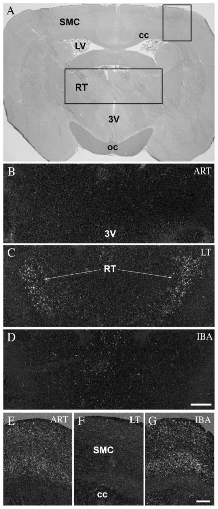
Fig. 4.
c-Fos expression in the reticular thalamic nucleus and the somatomotor cortex during the hibernation bout. A: Coronal section of the brain of a 13-lined ground squirrel 1.5 mm posterior to the level of the bregma. Insert areas (1, 2) are shown enlarged in B–D and E–G, respectively. The reticular thalamic nucleus showed no activity in awake animals (ART and IBA) (B,D), meanwhile during the torpor phase of the hibernation bout (LT), strong c-fos expression was detected in the nucleus (C). In contrast, activated neurons were found in the somatomotor cortex in only awake animals (ART and IBA) (E,G), but not during torpor (LT) (F). 3V, third ventricle; cc, corpus callosum; LV, lateral ventricle; oc, optic chiasm; RT, reticular thalamic nucleus; SMC, somatomotor cortex. Scale bars = 100 μm in B–D; 50 μm in E–G.

