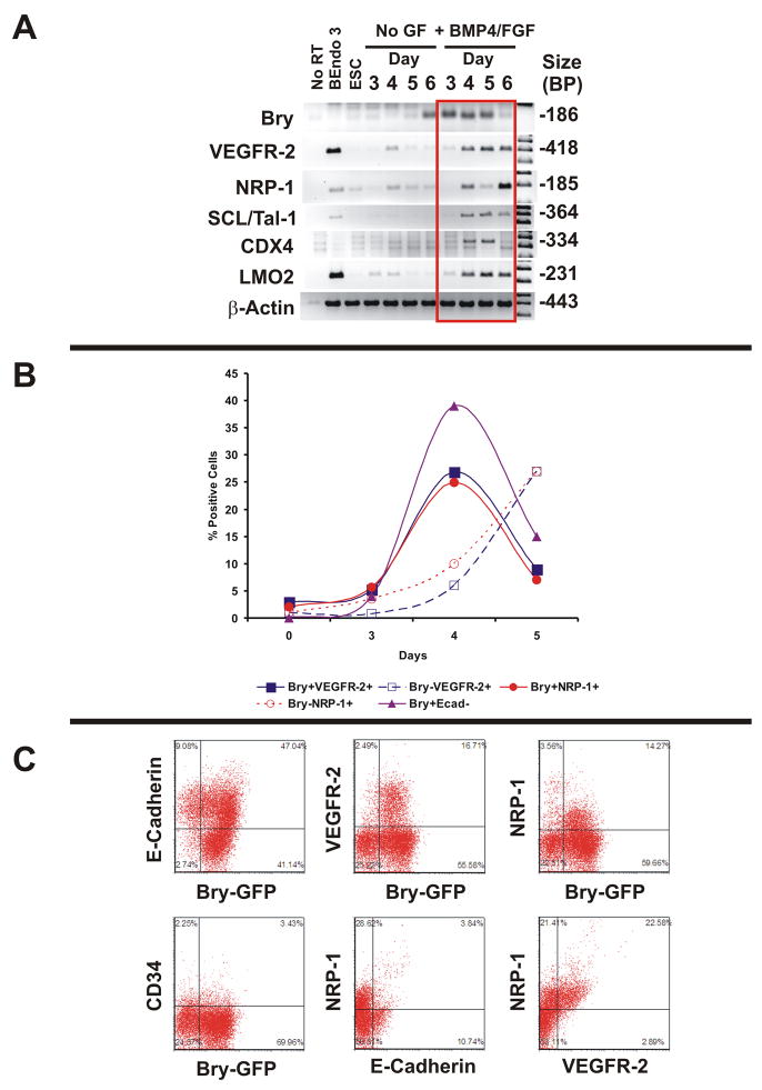
Figure 1. NRP-1 Expression Coincides With Bry and VEGFR-2 in Differentiating Murine ESCs.
Panel A: RT-PCR Time course analysis of embryoid bodies from Bry GFP murine ESCs in the absence and presence of BMP4 and bFGF. Results are representative of 3 experiments. The murine endothelialioma cell line bEnd.3: positive control for VEGFR-2, NRP-1, SCL/Tal-1, and LMO 2.
Panel B: Time course analysis of murine ESC differentiation as embryoid bodies in serum free conditions. Percent positive cells determined by flow cytometry analysis
Panel C: Representative flow cytometry plots of Bry GFP murine ESCs stained with antibodies to E-Cadherin, VEGFR-2, NRP-1, and CD34. Double staining experiments with NRP-1/E-cadherin and NRP-1/VEGFR-2 were performed in day 4 embryoid bodies in Rosa 26 mESCs. Quadrants set with IgG control. Data are representative of 5 experiments.

