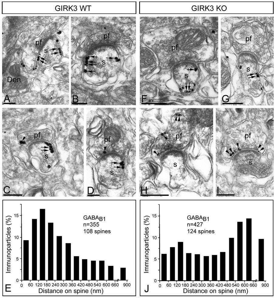
Figure 5. Subcellular regulation of GABAB receptors in GIRK3 KO mice.
Electron micrographs show the subcellular localization of GABAB1 in WT and GIRK3 KO mice, as revealed using a pre-embedding immunogold method. (A–D) In the WT, immunoparticles for GABAB1 were mainly localized along the extrasynaptic plasma membrane of PC spines (s) (arrows), but close to the glutamate release site, as well as at the edge of PSDs (crossed arrows) of PC spines (s). At the presynaptic level, few immunoparticles for GABAB1 was found in parallel fibre terminals (pf) (arrowheads). (F–I) In the GIRK3 KO cerebellum, immunoparticles for GABAB1 were also localized along the extrasynaptic plasma membrane of PC spines (s) (arrows), although most of them far away from the glutamate release site. At presynaptic sites, an increase in the number of immunoparticles for GABAB1 was detected in parallel fibre terminals (pf) (arrowheads). (E,J) Distribution of immunoreactive GABAB1 in relation to glutamate release sites in PC dendritic spines of WT and GIRK3 KO mice, respectively, as assessed from immunogold reactions. Immunoparticles were recorded in 60-nm-wide bins along the extrasynaptic plasma membrane of PC spines. Data are expressed as the proportion of immunoparticles at a given distance from the edge of the synaptic specialization. The measurements show that GABAB1 is redistributed along the extrasynaptic plasma membrane of PC spines in the GIRK3 KO mice. Scale bars, 0.2 µm.

