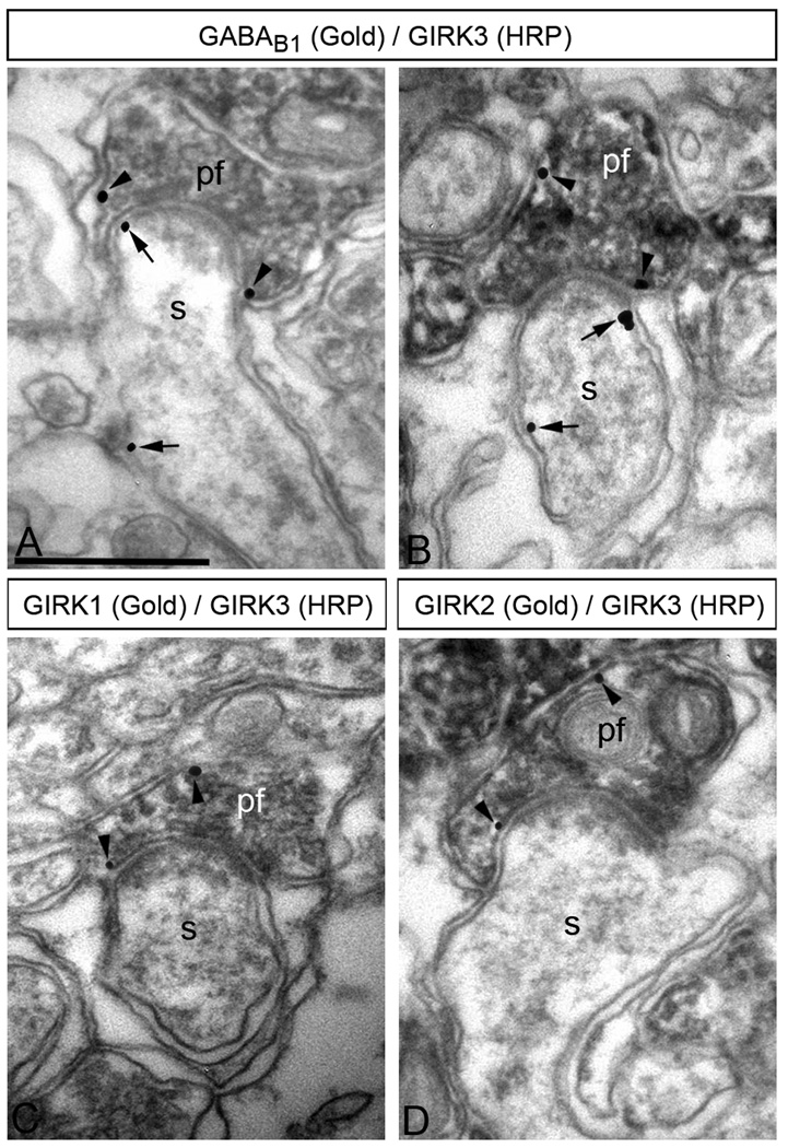
Figure 6. GABAB receptors co-localize with GIRK subunits in parallel fibre terminals.
Electron micrographs show co-localization of GABAB1 and GIRK3 (A,B), as well as co-localization of GIRK1 or GIRK2 with GIRK3 (C, D), in the same parallel fibre terminal, as revealed using double-labelling pre-embedding methods. (A–B) The peroxidase reaction product (HRP) indicating GIRK3 immunoreactivity filled parallel fibre terminals (pf), whereas immunoparticles (GABAB1 immunoreactivity) were mainly located along the extrasynaptic plasma membrane, and occasionally in the presynaptic membrane specialization of parallel fibre terminals (pf) establishing excitatory synapses on PC spines (s) (arrows). (C,D) The peroxidase reaction product (GIRK3 immunoreactivity) filled parallel fibre terminals (pf), whereas immunoparticles (GIRK1 immunoreactivity or GIRK2 immunoreactivity) were mainly located along the extrasynaptic plasma membrane, and occasionally in the presynaptic membrane specialization of parallel fibre terminals (pf) establishing putative excitatory synapses on PC spines (s) (arrows). Scale bars, 0.5 µm.

