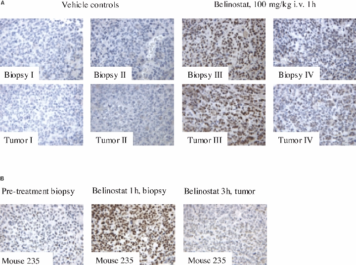
Fig. 4.

Acetylation of H4 in A2780 xenograft mice before and after belinostat treatment. (A) Acetylated H4 in four biopsies and their representative tumors (I–IV) (Experiment 2). No (A) or weak (IZI) H4 acetylation is observed in vehicle control mice, whereas strong (III) or moderate (IV) H4 acetylation is observed 1 h after intravenous treatment with belinostat (100 mg/kg) in both biopsies and tumors. (B) H4 acetylation in vivo in biopsies and tumor from the same mouse during belinostat treatment (100 mg/kg i.v.) (Experiment 3). The pretreatment biopsy shows weak H4 acetylation, whereas strong H4 acetylation is seen 1 h after treatment. After 3 h, the H4 acetylation is weak and has decreased to a level similar to that in the pretreatment biopsy.
