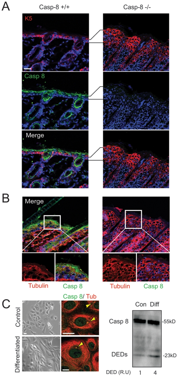
Figure 1. Effects of caspase-8 downregulation in skin maturation.
A. Immunohistochemistry analysis performed in skin from wildtype and caspase-8 knock out mice. Caspase-8 DEDs are stained in green, keratin 5 in red and nuclei in blue. Brackets denote expanded epidermis in the caspase-8 knockout mice. B. Microtubule staining (red channel) in skin from wild type and caspase-8 knockout mice. Insets show magnified views of the microtubule patterning in wild type and caspase-8 knockout mice. C. Left, Confocal images of HaCat undifferentiated and differentiated cells stained for caspase-8 DEDs (green channel) and microtubules (red channel). Yellow arrows indicate centrosomes. Right, Immunoblot analysis of caspase-8 expression in undifferentiated or differentiated keratinocytes. Lysates were probed with antibody recognizing the DEDs of caspase-8.

