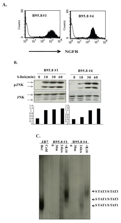
Figure 2. LMP1 induces STAT1/3/DNA complexes 24 hr after activation.
A. BL41 clones expressing chimeric NGFR-LMP1 B95.8 were created as described in Materials and Methods and are referred to as B95.8 #1 and B95.8 #4. Expression of NGFR-LMP1 on the surface of BL41 cells was tested by flow cytometry using anti-NGFR-biotinylated antibodies and streptavidin-PE. Filled histogram shows NGFR staining in BL41 clones and thin line histogram indicates NGFR background staining in the untransfected parental BL41 line. B. B95.8 #1 and B95.8 #4 clones were incubated with anti-NGFR and goat anti mouse IgG for 0, 10, 30 or 60 minutes. The cells were harvested, lysed in phospholysis buffer and resolved on SDS-PAGE. After transfer to nitrocellulose, anti-phospho-JNK and anti-JNK antibodies were used for Western blot. Densitometry analysis is shown below the blots and represents the ratio of pJNK to total JNK. C. NGFR-LMP1 BL41 clones were crosslinked with anti-NGFR and goat anti-mouse Ig for 0, 30 minutes or 24 hours or treated with 10 ng/ml of recombinant human IL-10 for 30 minutes. 40 μg of whole cell lysates of BL41 clones and JB7 SLCL were incubated with 32P labelled hSIE probe with/without 1000x excess of cold probe and resolved on 4% non-denaturing acrylamide gel followed by autoradiography. Data are representative of three or more independent experiments.

