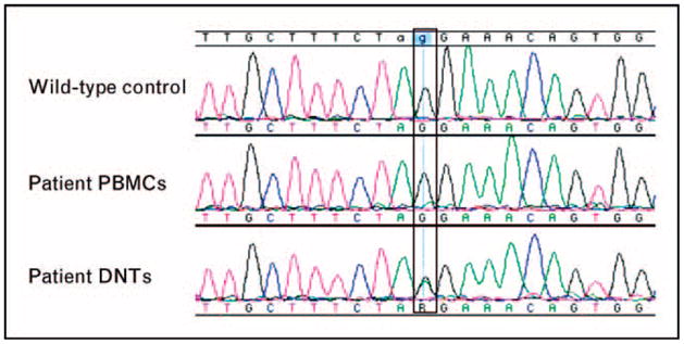
Figure 5. Alignment of genomic DNA sequencing data at the Fas exon 8 acceptor splice site: wild-type control (top), patient peripheral blood mononuclear cells (middle), and patient purified double-negative α/β T-cell receptor-positive T cells (bottom).

The mutant peak in the patient’s PBMCs is not distinguishable from baseline noise, and the mutation is only detectable in the chromatogram for the isolated DNTs. DNTs, double-negative α/β T cells; PBMCs, peripheral blood mononuclear cells.
