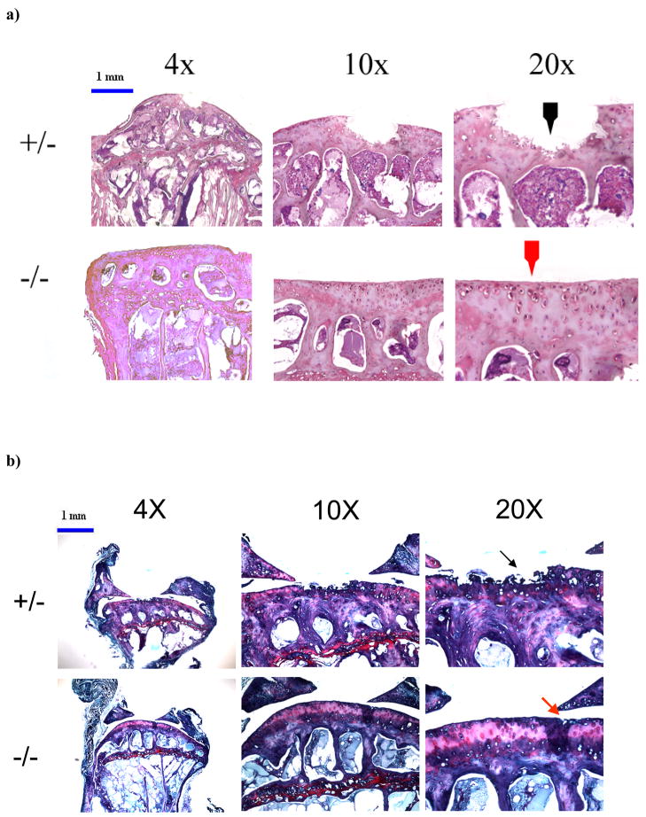
Figure 4. Defects in the articular cartilage of knee joints in adult mice.
a) Micrographs of H&E histology showing damage at the articular surface in knee joints of DN p38 (+/−) mice (black arrow) and a corresponding region in WT (−/−) mice (red arrow). Bar = 1mm.
b) Micrographs of Safranin-O staining showing greater damage to the articular cartilage in knee joints of DN p38 (+/−) mice (black arrow) compared to WT (−/−) mice (red arrow). Bar = 1mm.

