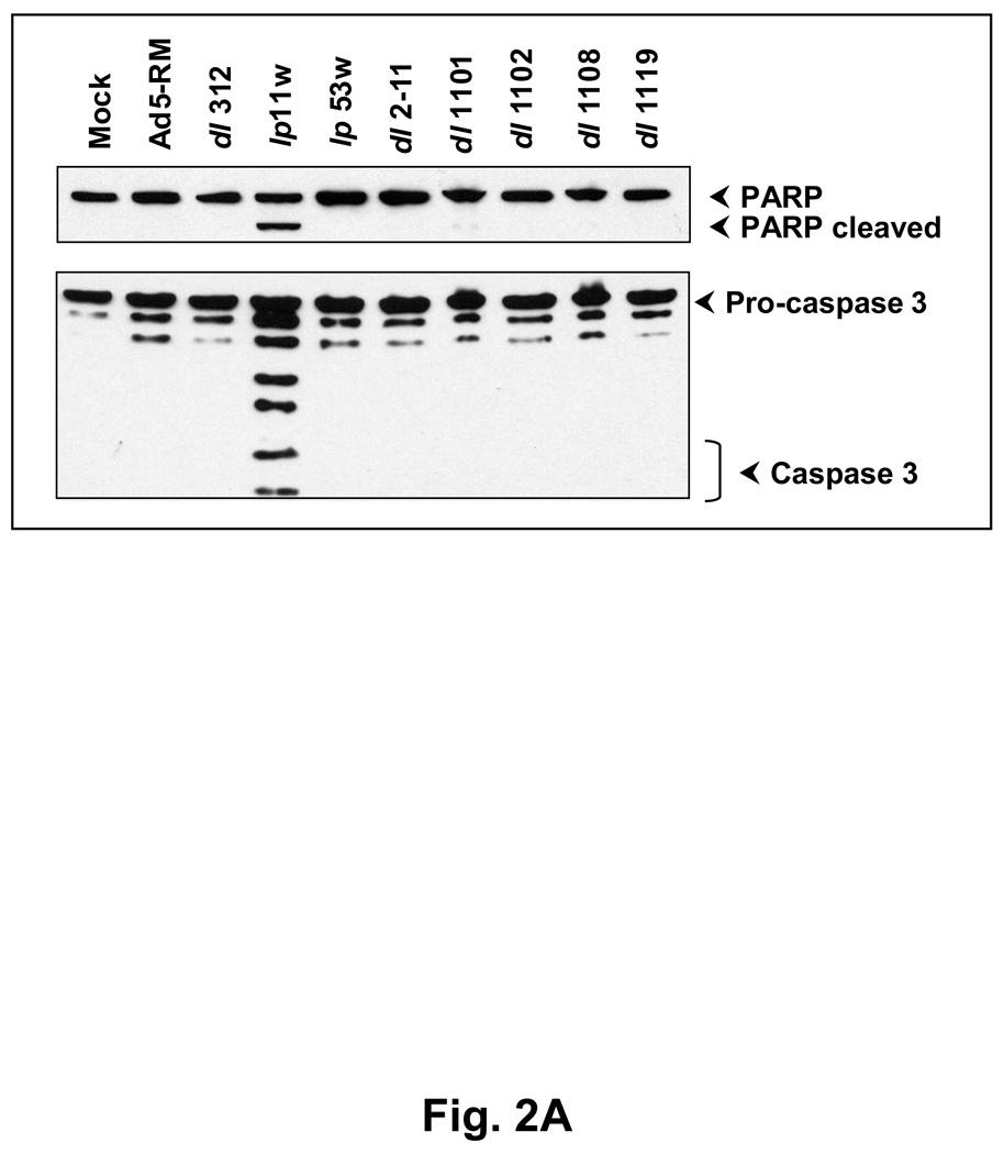
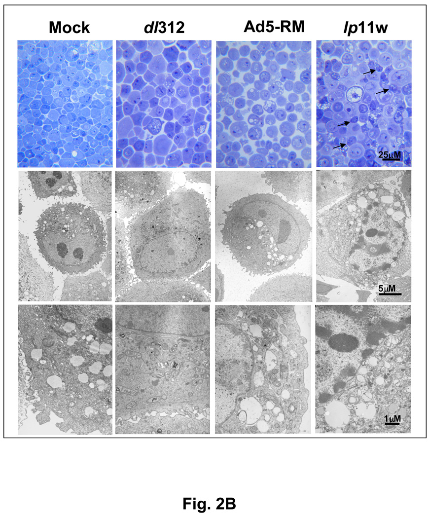
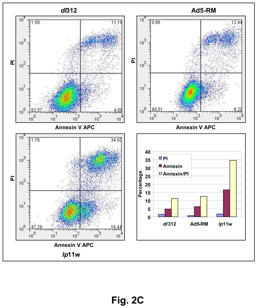
Figure 2. Apoptotic activity of lp11w.
(A). Effect of Ad5 mutants on caspase activation. SCC25 cells were infected with indicated mutants and proteolytic processing of pro-caspase 3 and PARP were determined by western blot analysis. (B). Ultra-structure of infected cells. Thin sections of Ad-infected cells were analyzed by light microscopy (top panel) and by electron microscopy (bottom two panels). Apoptotic cells in the top panel of lp11w-infected samples are pointed out by arrows. (C). Flow cytometric analysis of annexin V-staining patterns of Ad-infected cells. The quantification of annexin V-positive and propidium iodide-positive cells is shown in the bar diagram.


