Figure 1.
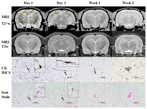
In vivo cellular MRI with histological validation of brain metastases in group 1 rats. Representative group 1 rats that received 3 × 10⁶ FEPro labeled 231BRL cells with each column matched to the same animal. T2*-weighted images demonstrate diffuse brain metastasis of tumor cells as hypointense voxels on days 1 and 3 post intracardiac injection. Arrowheads mark some of the hypointense regions. Growing metastatic breast cancers were greater than 200-300 μm in size at week 2. T2-weighted image shows hyperintense tumor at left hippocampus at week 2 (arrowhead). Cytokeratin immunohistochemical staining (CK IHCS) of the brain showed tumor cells (i.e., brown) in the microvasculature of the brain at the early period (day 1-3, arrow) and growing mass at the later time points (week 1-2). Prussian blue iron staining were compatible findings to CK IHCS staining for tumor cells. Bar: MRI = 4 mm, histology = 200 μm.

