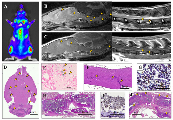
Figure 4.
Central Nervous System and skeletal involvement by breast cancer. A) Bioluminescence image of rat at week 3 shows high photon flux activity from the brain, spine, and joints. (B) Sagittal T2w MRI and (C) contrast enhanced T1w MRI show hyperintense lesions on the brain, spinal cord, and vertebral bodies (arrowheads). Histological section of brain (D) and spinal cord (F) with hematoxylin and eosin (HE) staining from group 2 rat euthanized at week 4 reveals numerous metastases (arrowheads). E) Prussian blue staining of the consecutive brain section from D shows few isolated iron positive cells near the tumor (arrows). H) Thoracic spine with tumor infiltration on HE stain. Cytokeratin immuno-histochemical staining of the bone marrow aspirates (G) and Spine (I) is positive for tumor. J) Knee joint metastasis with extraskeletal involvement is seen (arrowheads) on HE stain.

