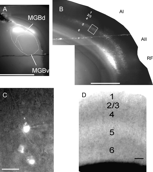
Figure 1.
Fluorescent (Zeiss filter set #15: Excitation filter BP 546/12, Emission filter LP 590, Dichroic FT 580) photos, taken as monochrome and of a slice while in the recording apparatus. (A) The ×5 photograph of typical injection site in the medial geniculate body. MGBd = dorsal division of the MGB. MGBv = ventral division of the medial geniculate body. (B) Retrograde cortical labeling in the same slice. Note that the MGBd injection produces retrogradely labeled neurons in layers 5 and 6 of the auditory cortex, with a predominance of layer 5 label in the primary auditory cortex (AI) and both layers 5 and 6 of the secondary auditory cortex (AII). RF = rhinal fissure. Scale bar for (A, B) = 1mm. (C) The ×40 view of labeled cells from dotted box in (B). A patch pipette is shown patched onto the top cell. Scale bar = 25 μm. (D) The ×10 IR-DIC photograph of the primary auditory cortex while in the recording chamber to illustrate the lamination patterns used to designate individual layers in this study. Scale bar = 100 μm.

