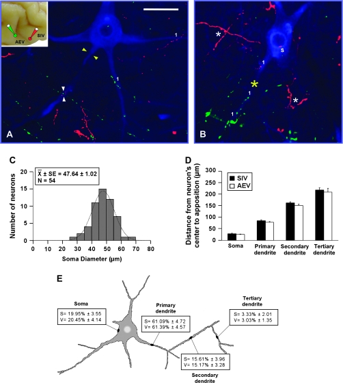
Figure 8.
Modality-convergence pattern of AEV–SIV onto SMI-32 immunostained neurons. Anterogradely labeled fibers from AEV (green) and SIV (red) were observed either across the different compartments (A) or within a single compartment (yellow asterisk in B) of the SMI-32 immunostained neuron. Appositions distributed on the soma (“s”) as well as on primary (“1”), secondary (“2”), and tertiary (“3”) dendrites of SMI-32 immunostained neurons (A, B). The soma diameters of neurons receiving convergent inputs from AEV and SIV are shown in (C). Appositions from AEV and SIV were located at similar distances on the cell body as well as on dendrites (D), and the highest percentage of appositions was observed on primary dendrites and less frequently on the other neuron's compartments (E). No significant differences were observed either in the distribution or in the percentage of appositions from AEV–SIV onto the SMI-32 immunostained neuron's compartments. Scale bar = 50 μm in (A).

