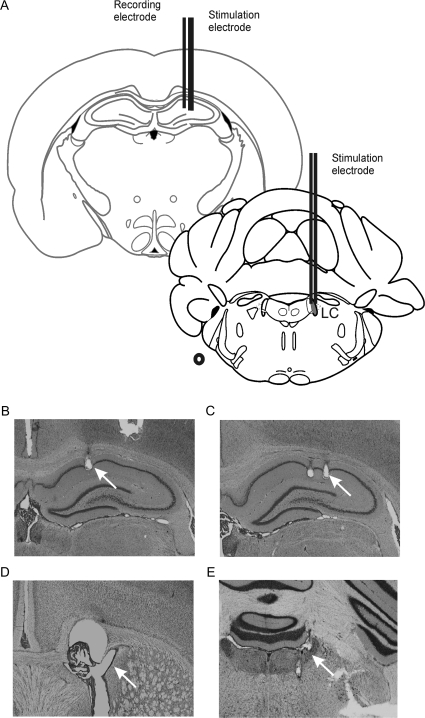
Figure 2.
Placement of electrodes and cannula. (A) Schematic representation of electrode placement in the hippocampus and LC (redrawn from Paxinos and Watson's Atlas, 1997). (B) Microphotograph showing recording electrode placement in the stratum radiatum of CA1. Microphotograph of bipolar stimulation electrode placement in (C) the SC input from CA3 to CA1 and the (E) LC. (D) A cannula was inserted into the lateral cerebral ventricle to enable drug or vehicle solution administration.

