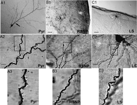
Figure 10.
Three physiologically identified spiny neurons within the PPC. DAB-stained photomicrographs from 3 PPC neuron cell types: a layer II PYR (A), layer III RSNP (B), and layer I LS (C) cells were obtained with a light microscope using a ×20 objective (A1, B1, C1) or a ×100 oil immersion objective (A2, B2, C2), with the rectangle in these latter panels indicating the area of dendrites highlighted in the final panel (A3, B3, C3). Scale bar = 50 μm in (A1, B1, C1). Scale bar = 10 μm in (A2, B2, C2). Note the large apical dendritic trunk indicated by the black arrow (A1) and the high density of dendritic spines (A3) found on PYR cells.

