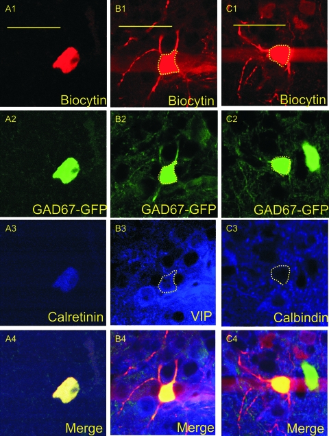
Figure 13.
Confocal microscopy images of immunopositive interneurons. Images were obtained using a ×60 objective and represent a single cell that was triple stained for neurobiotin (A1, B1, C1), GFP (A2, B2, C2), and CR (A3), VIP (B3), or calbindin (C3). The final panels represent the above 3 images merged together (A4, B4, C4). (A1–A4) are from an IS cell found in layer I-B of the PPC, (B1–B4) are from an RSNP cell found in layer II, and (C1–C4) are from an RSNP cell in layer II that was found to show no immunoreactivity for calbindin. Scale bar = 50 μm.

