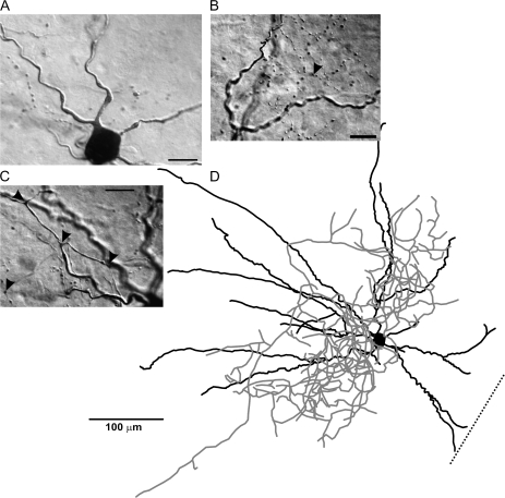
Figure 8.
FS interneuron of the PPC. A typical example of a Neurolucida reconstruction of a FS cell (D). The dendrites of FS cells were typically aspiny or smooth in appearance (A), and the axons typically showed numerous boutons (A, B) and enlargements at branching points (C). The spiking patterns for this cell are represented in Figure 1D. Scale bar in panel (D) = 100 μm. Scale bars in panels (A, B, C) = 15 μm.

