
Figure 6.
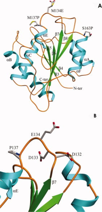
(A) Ribbon diagram of lipase mutant 4D3, indicating the relative position of three mutations. (B) Location of E134, which is adjacent to two negatively charged residues, D132 and D133. [Color figure can be viewed in the online issue, which is available at www.interscience.wiley.com.]