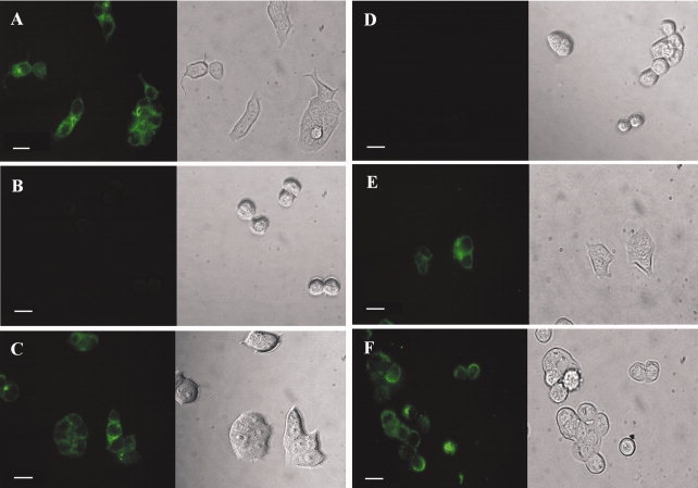
Figure 3.

Fluorescence images of MCF7 and HT-29 cells. The cells were treated with 0.1 nM of dsFv-B3-R8C, E8C-LLO, and B3-LLO, respectively. All proteins were labeled with fluorescein. (A) MCF7 cells, positive for the antigen Lewis Y, exhibited green fluorescence after binding of the antibody fragment dsFv-B3-R8C. (B) HT-29 cells, negative for Lewis Y, did not fluoresce after incubation with dsFv-B3-R8C. (C) MCF7 cells also showed green fluorescence due to the binding of the immunotoxin B3-LLO, whereas HT-29 did not (D). The isolated cytolysin E8C-LLO interacted with both MCF7 cells (E) and HT-29 cells (F). On the left side there are fluorescence images, the right side shows the corresponding images from transmitted light. The length of the bar is equivalent to 20 μm.
