
Fig. 2.
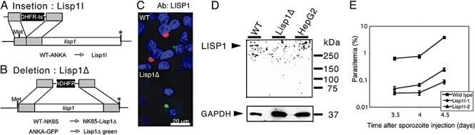
Targeted gene disruption of lisp1. A. Schematic representation of Lisp1I gene disruption. B. Schema of Lisp1Δ gene disruption. Shaded boxes indicate the regions of homology used for double-cross-over recombination; black boxes indicate the selectable marker; Met and the asterisk (*) indicate the initiation and stop codons respectively. C. Absence of LISP1 protein in Lisp1Δ parasites. Microscopy image of HepG2 cells 48 h post infection with WTGreen or Lisp1ΔGreen. LS were labelled with anti-LISP1 antibody and Alexa 647-conjugated secondary antibody (red); nuclei stained with DAPI (blue). D. Western blot analysis of extracts of HepG2 cells 48 h post infection with ANKA (WT) or Lisp1ΔGreen sporozoites. Arrowheads indicate the bands of LISP1 and human GAPDH. E. Lisp1I sporozoites have a decreased infectivity in the mammalian host. A total of 30 000 wild-type or Lisp1I sporozoites, from two independent Lisp1I clones, were injected intravenously into rats and blood parasitaemias were examined by Geimsa staining. The error bars show the standard errors from three rats.