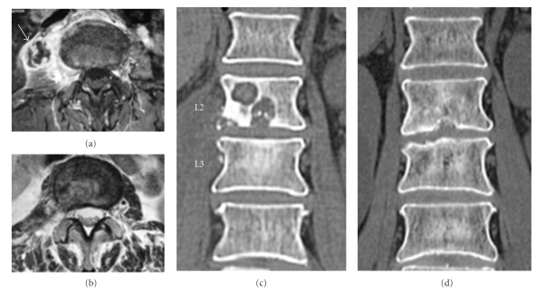
Figure 2.
(a) Preoperative gadolinium imaging, axial image. Psoas abscess formation is visible (arrow), along with rim enhancement. (b) T2-weighted imaging at 3 months postoperatively, axial image. The iliopsoas abscess has disappeared. (c) Preoperative CT shows clear cavitations within the L2 vertebral body. (d) CT at 12 months postoperatively indicates that the cavity within the vertebral body has disappeared and has been remodeled.

