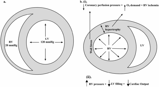
Fig. (1).
Short axis view of the right (RV) and left (LV) ventricles in a patient with normal (a) pulmonary pressures and in a patient with pulmonary hypertension (b). In normal individuals, note the crescent shape of the RV and the thin wall. In pulmonary hypertension, elevations in RV pressures increase myocardial wall stress. As the RV hypertrophies, myocardial oxygen demands also increase, culminating into a myocardial supply/demand mismatch (i). With increased RV filling pressure and volume, the septum is displaced towards the LV, reducing cardiac output (ii). Reproduced with permission from Chin et al. [4].

