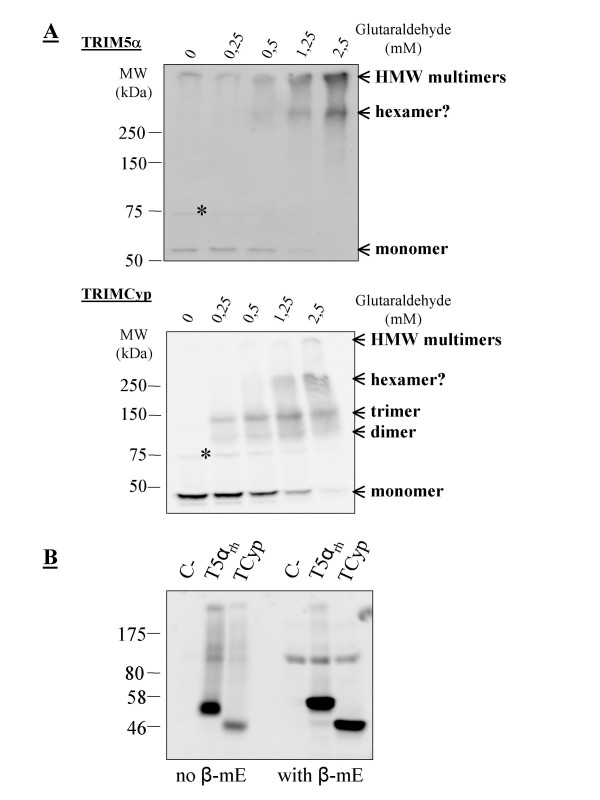
Figure 1.
Multimerization profiles of TRIM5α and TRIMCyp. A, 0.5% NP40 lysates were prepared from Mus dunni tail fibroblast cells (MDTFs) stably expressing FLAG-tagged TRIM5αrh or owl monkey TRIMCyp. The soluble fraction of each lysate was divided in aliquots that were treated for 5 min with the indicated glutaraldehyde concentrations before proteins were denatured by boiling in the presence of SDS. Proteins were then separated on an 8% polyacrylamide gel, transferred to a nitrocellulose membrane, and probed with a rabbit anti-FLAG antibody (Cell Signaling). The apparent multimeric states are indicated on the right as deduced from the size of the bands. The star indicates an unspecific protein cross-detected by the FLAG antibody. B, Lysates were prepared from HeLa cells stably transduced with the same constructs as above and in the absence or presence of 100 μM β-mercaptoethanol as indicated.

