Figure 2.
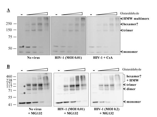
Multimerization of TRIMCyp in cells infected by HIV-1. A, MDTF-TRIMCyp cells were challenged with an HIV-1 vector expressing GFP, at a dose leading to infection of about 1% of the cells and either in the presence or not in the presence of 5 μM cyclosporine A (Sigma). After 6 hours of infection, cells were lysed in presence of increasing glutaraldehyde concentrations as in Fig. 1. Western blot analysis of FLAG-tagged proteins was performed as above. B, the experiment was repeated in the presence of 1 μM MG132 (Sigma) and using two different virus doses. The stars indicate cellular proteins cross-detected by the FLAG antibody as evidenced by analysis of lysates from parental cells (not shown).

