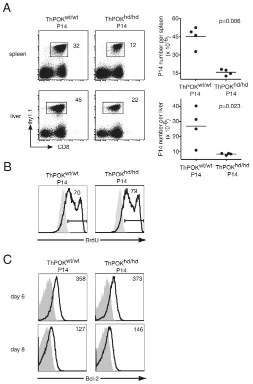
FIGURE 3.
Reduced accumulation of effector P14 T cells in the absence of functional ThPOK. A, CD44lowCD4−ThPOKhd/hd or ThPOKwt/wt P14 cells (Thy1.1) (104) were transferred into B6 hosts that were subsequently immunized with LCMV. At 8 days postinfection, the fraction of both types of P14 T cells and their absolute numbers in spleen and liver were determined. The results are representative of eight independent experiments for spleen and two independent experiments for liver. B, Host mice were injected with 1 mg of BrdU i.p. (open histograms) or left untreated (shaded histograms) at 6 days postinfection. Six hours later, cells harvested from the spleen were analyzed for BrdU incorporation. The numbers in the histograms are the percentages of BrdU+ cells among P14 cells. C, At the indicated time points the levels of Bcl-2 expression (open histograms) in ThPOKhd/hd P14 or ThPOKwt/wt P14 cells were determined by flow cytometry. Shaded histograms represent isotype controls and numbers refer to the MFI of Bcl-2.

