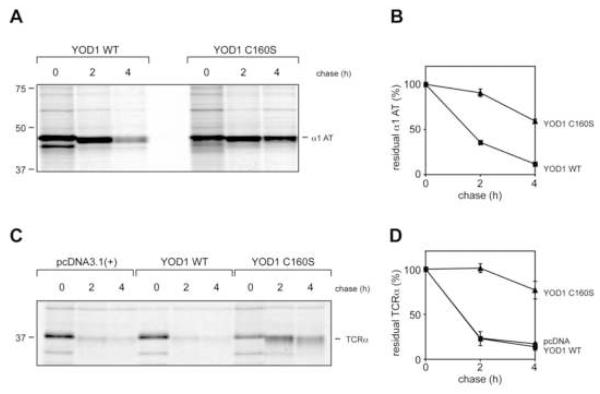
Figure 5. YOD1 C160S impairs dislocation of α1-antitrypsin and TCRα chain.
(A) 293T were cells transfected with α1-antitrypsin NHK (α1 AT), and YOD1 WT or YOD1 C160S, were pulse-labeled with 35S for 10 min and chased for the indicated time points. The cells were lysed in SDS and the lysates were immunoprecipitated with anti-α1-AT antibodies. The eluates were separated by SDS PAGE (12%) and the bands were visualized by autoradiography.
(B) Quantification of NHK levels. Plotted is the mean value of three independent experiments with the error bar corresponding to the standard deviation.
(C) 293T cells were transfected with TCRα, and either empty vector (pcDNA), YOD1 WT or YOD1 C160S. The pulse-chase experiment was performed as in Fig. 3 A. TCRα was retrieved from SDS-lysates by immunoprecipitation with anti-TCRα antibodies and visualized by autoradiography.
(D) Quantification of TCRα levels. Plotted is the mean value of two independent experiments with error bars.

